Cervical Spinal Stenosis
Introduction
Cervical stenosis is an abnormal narrowing of the spinal canal, which is the passageway for the spinal cord and nerve roots as they pass through the cervical spine. Cervical stenosis is most commonly due to degenerative changes which accumulate over the years including dic herniations, thickened ligaments and bony spurs. Patients often complain of neck and arm pain, numbness, tingling and sometimes weakness and balance difficulties. Most patients respond to non operative management while those with progressive lesions or neurologc injuries may require surgical treatment.
Causes
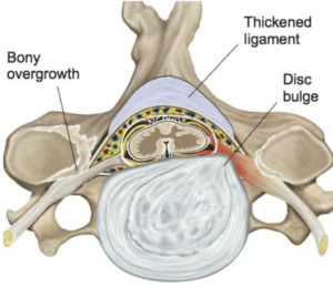
Most cases of cervical stenosis are related to degenerative changes of the cervical spine joints (disc and bilateral facet joints) which accumulate over the years. These include disc protrusions/herniations, thickened ligaments, and enlarged facet joints and bony spurs (figure 1). Instability in the form of spondylolisthesis can further narrow the spinal canal diameter. Other less common causes include trauma (fractures), infections, congenital abnormalities, and tumors.
Figure 1
Illustration demonstrating cervical stenosis due to thickened ligamentum flavum, bony spur, and disc bulge/protrusion with compression of the spinal cord and left cervical nerve root.

Diagnosis
The diagnosis of cervical stenosis is made following a detailed history and physical examination by an experienced spine surgeon and confirmed with appropriate imaging studies. Patients with stenosis compressing a nerve root frequently describe arm pain, numbness, tingling and or weakness (cervical radiculopathy); patients with stenosis compressing the spinal cord may describe bilateral arm pain, intrinsic hand weakness (dropping objects, difficulty opening jars), numbness (difficulty with fine objects and jewelry), lower extremity weakness or balance problems (cervical myelopathy). Some patients have both:myeloradiculopathy. Physical exam findings include decreased neck motion with pain, especially when looking up, numbness, weakness, poor balance, and abnormal reflexes. Clinical diagnosis is confirmed with imaging studies especially MRI which best demonstrates spinal anatomy and any correlating neurologic compression (figure 2). The MRI is non invasive and does not involve radiation (x-ray energy). Some patients are unable to have an MRI due to presence of a pacemaker or other metallic (iron based) loose bodies. In these cases a CT myelogram, which involves injecting a contrast dye into the spinal canal to outline the nerves, will provide good visualization of the spine and stenotic lesions
Figure 2
A sagittal section (looking from the side) image of the cervical spine in the midline with MRI demonstrating cervical stenosis and compression of the spinal cord. Arrows demonstrate herniated discs from the front (anteriorly) and the thickened ligaments in the back (posteriorly) “pinching” the spinal cord. Notice the complete obliteration of the fluid filled epidural space producing a severe stenosis at C5/6 and moderate at C4/5.

Non operative treatment
Most patients with mild to moderate cervical stenosis are able to manage their condition with nonoperative treatment. This begins with activity modification to avoid activities that aggravate and inflame the “arthritis” including extension based activities. A soft collar can provide some support with activities that aggravate myeloradicular symptoms. NSAIDs and possible steroid injections including epidural steroids can decrease pain and inflammation and facilitate rehabilitative efforts. Patients with severe pain my require temporary use of narcotics (oxycodone, tramadol, hydrocodone) or muscle relaxants in the presence of muscle spasm. Once the pain and neurologic symptoms improve physical therapy (PT) is very helpful to improve neck motion and strength and increase activities and functional abilities. Therapy also helps educate patients regarding proper body mechanics and posture. PT may also other modalities including ice, heat, ultrasound, TENs unit, and massage. Patients are then instructed in developing a home exercise program that includes core range of motion and strengthening exercises as well as low impact aerobic exercise (swimming, water aerobics, stationary bike, elliptical, etc).
Patients with chronic nerve pain can be treated with “anticonvulsant” medications including gabapentin (Neurontin), pregabalin (Lyrica), topiramate (Topamax) which are thought to act by preventing abnormal increases in brain activity (usually through Na or Ca channels).
Antidepressants such as nortriptyline and duloxetine (Cymbalta) may also be helpful for patients with chronic pain by modulating pain transmission in the central nervous system.
Patients should be educated on the natural history of cervical stenosis, the probability of lesions progressing over time, and the risk for neurologic injury, even paralysis, with progression or trauma. Patients should be re-evaluated on a regular basis, implement fall precautions, and undergoe repeat imaging if new neurologic changes develop.
Operative Treatment
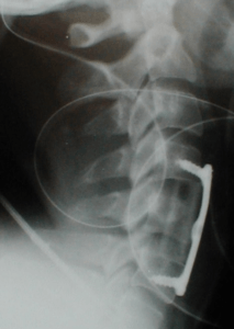
Patients with severe stenosis, myelopathy or significant neurologic deficit, or who fail to improve with non operative treatment may require surgical management. Surgical treatment involves removal of the stenotic lesion. Disc herniations are treated by an anterior discectomy to decompress the nerves and or spianl cord and fusion and instrumentation to stabilize the spine, ACDFI (figure 3).
Figure 3
Patient from figure 2 with myeloradiculpathy treated with cord decompression via C5 corpectomy, fusion with fibular strut allograft, and plate fixation C4-6.

Posterior lesions are treated with laminectomy, laminoplasty, or laminectomy with posterior cervical fusion and instrumentation if instability is present. Minimally invasive spine surgery (MISS) is an option in smaller lesions involving less severe stenosis and open approaches for more involved cases. Surgery for spinal stenosis is one of the most successful orthopaedic procedures with good to excellent results in the majority (>90%) of patients. Outcome studies demonstrate similar quality of life (QOL) scores following spinal decompression compared to total hip and knee arthroplasty and superior cost utility (QALY).
